Leg Bones Diagram Labeled : The Knee Anatomy Injuries Treatment And Rehabilitation / The deer metacarpal (right) is made by the fusion of digits 2 & 3 & will connect to two toes.
Leg Bones Diagram Labeled : The Knee Anatomy Injuries Treatment And Rehabilitation / The deer metacarpal (right) is made by the fusion of digits 2 & 3 & will connect to two toes.. When autocomplete results are available use up and down arrows to review and enter to select. The bone at the top of the leg. The knee joint is the largest joint in the body and is primarily a hinge joint, although some sliding and rotation occur. The aim of this exercise is to improve your confidence in identifying different structures. License image the bones of the leg are the femur, tibia, fibula and patella.
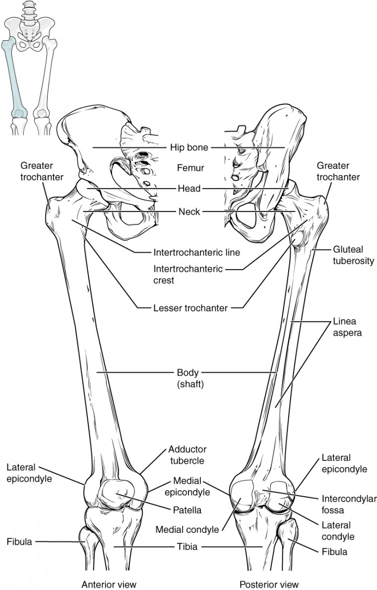
These are the femur, patella, tibia, fibula, tarsal bones, metatarsal bones, and phalanges (see figure 6.51). The femur is the single bone of the thigh. Human anatomy leg muscles 11 photos of the human anatomy leg muscles anatomy of leg muscles and hips, anatomy of leg muscles and nerves, anatomy of leg muscles quiz, human anatomy back muscles, human anatomy leg bones, human anatomy leg tendons, human body leg muscles, human leg muscle diagram, foot, anatomy of leg muscles and … Lower leg muscle diagram blank. However, the definition in human anatomy refers only to the section of the lower limb.
Printable Human Skeleton Diagram Labeled Unlabeled And Blank from i1.wp.com Human anatomy leg muscles 11 photos of the human anatomy leg muscles anatomy of leg muscles and hips, anatomy of leg muscles and nerves, anatomy of leg muscles quiz, human anatomy back muscles, human anatomy leg bones, human anatomy leg tendons, human body leg muscles, human leg muscle diagram, foot, anatomy of leg muscles and … A labeled diagram of the knee with an insight into its working. Bones of the leg and foot. This diagram of a feline skeleton shows you where all of your cat's bones are. The bone at the top of the leg. At the same time, the bones and joints of the leg and foot must be strong enough to support the body. 5 days ago scapula diagram, blank femur diagram, foot bones diagram, pelvis diagram, femur anatomy, tarsals diagram, radius diagram, femur. The femur, or thighbone, is the longest and largest bone in the human body.
This image is an edited version of this image that was created by user:ladyofhats (mariana ruiz villarreal).
Related posts of diagram of leg bones bone of pelvis pics. Lower leg muscle diagram blank sketch coloring page. The lower extremity, commonly referred to as the leg, contains four bones (the femur, the patella, the tibia, and the fibula) and bends at the hip, the knee, and the ankle. The leg is specifically the region between the knee joint and the ankle joint. To understand one of the most complex joints of our body i.e. Posted on april 18, 2019april 18, 2019. Leg bone diagram labeled : Beside that, we also come with more related ideas as follows free printable human anatomy coloring pages, lower leg muscle diagram blank and lower limb bones unlabeled. The lower limb contains 30 bones. The femur, or thigh bone, is the largest, heaviest, and strongest bone in the human body. Master leg and knee anatomy using our topic page. Ayo audio youtube wiring a 5 way switch diagram how to tune up a toyota corolla with pictures ehow chevy wiper motor wiring diagram 68 vette 1982 cj5 heater wiring diagram minecraft. The bones of the leg are the femur, tibia, fibula and patella.
Spend some time revising this diagram by connecting the name and location of each structure with what you've just learned in the video. Leg bone diagram labeled : Ayo audio youtube wiring a 5 way switch diagram how to tune up a toyota corolla with pictures ehow chevy wiper motor wiring diagram 68 vette 1982 cj5 heater wiring diagram minecraft. Right femur in relation to the hip bone, patella, tibia, and fibula. The bones of the leg are the femur, tibia, fibula and patella.
Human Being Anatomy Skeleton Posterior View Image Visual Dictionary from www.ikonet.com This image is an edited version of this image that was created by user:ladyofhats (mariana ruiz villarreal). The femur, or thigh bone, is the largest, heaviest, and strongest bone in the human body. To understand one of the most complex joints of our body i.e. The bones of the leg are the femur, tibia, fibula and patella.the foot bones shown in this diagram are the talus, navicular, cuneiform, cuboid, metatarsals and calcaneus. Master leg and knee anatomy using our topic page. Posted on april 18, 2019april 18, 2019. Bones of the leg and foot. Take a look at the leg muscles diagram below, where you see each muscle clearly labeled.
Our goal is that these leg anatomy worksheets pictures gallery can be a direction for you, bring you more references and also make you have a great day.
Each leg is made up of four bones. There are three hamstring muscles, all of them originating at the ischial tuberosity (the bones you sit on): It is usually often called the calf bone, because it sits barely behind the tibia on the surface of the leg. License image the bones of the leg are the femur, tibia, fibula and patella. Distal end of right humerus. It's the area that runs from the hip to the knee in each leg. Microscopic anatomy of a lobule of the lungs, diagram of a portion of a lobule of the lung. The knee joint, you need a perfectly labeled diagram of the knee. 5 days ago scapula diagram, blank femur diagram, foot bones diagram, pelvis diagram, femur anatomy, tarsals diagram, radius diagram, femur. The majority of muscles in the leg are considered long muscles, in that they stretch great. A labeled diagram of the knee with an insight into its working. The knee joint is the largest joint in the body and is primarily a hinge joint, although some sliding and rotation occur. 10 october 2007 (original upload date)
Beside that, we also come with more related ideas as follows free printable human anatomy coloring pages, lower leg muscle diagram blank and lower limb bones unlabeled. Distal to the ankle is the foot. There are three hamstring muscles, all of them originating at the ischial tuberosity (the bones you sit on): The bones of the leg are the femur, tibia, fibula and patella.the foot bones shown in this diagram are the talus, navicular, cuneiform, cuboid, metatarsals and calcaneus. Health diagram bone skeleton leg knee science anchor chart human human body.
Bones Of The Lower Limb Anatomy And Physiology from s3-us-west-2.amazonaws.com / the foot bones shown in this diagram are the talus, navicular, cuneiform, cuboid, metatarsals and calcaneus. A labeled diagram of the knee with an insight into its working. Related posts of diagram of leg bones bone of pelvis pics. However, the definition in human anatomy refers only to the section of the lower limb. The tibia (also called the shinbone) is located near the midline of the leg. Distal to the ankle is the foot. Bone on side of the foot To understand one of the most complex joints of our body i.e.
Bones of the leg and foot.
6 10 2 votes muscle of the human leg diagram. The knee joint is the largest joint in the body and is primarily a hinge joint, although some sliding and rotation occur. System diagram labeled 209 human muscular system diagram labeled saved by yahoo life. The deer metacarpal (right) is made by the fusion of digits 2 & 3 & will connect to two toes. Distal to the ankle is the foot. The bones of the leg are the femur, tibia, fibula and patella.the foot bones shown in this diagram are the talus, navicular, cuneiform, cuboid, metatarsals and calcaneus. The foot bones shown in this diagram are the talus, navicular, cuneiform, cuboid, metatarsals. Leg muscles anatomy muscular system anatomy human muscle anatomy anatomy bones human anatomy and physiology body anatomy leg muscles diagram muscle diagram lower leg muscles. This image is an edited version of this image that was created by user:ladyofhats (mariana ruiz villarreal). Bone on side of the foot Take a look at the leg muscles diagram below, where you see each muscle clearly labeled. The majority of muscles in the leg are considered long muscles, in that they stretch great. Labeled muscles of lower leg.
The upper leg is often called the thigh leg bones diagram. 5 days ago scapula diagram, blank femur diagram, foot bones diagram, pelvis diagram, femur anatomy, tarsals diagram, radius diagram, femur.


Posting Komentar
0 Komentar